


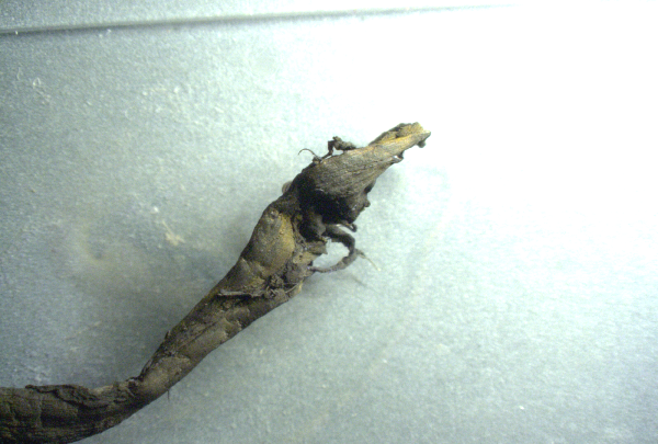
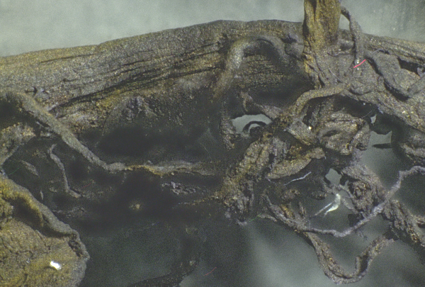
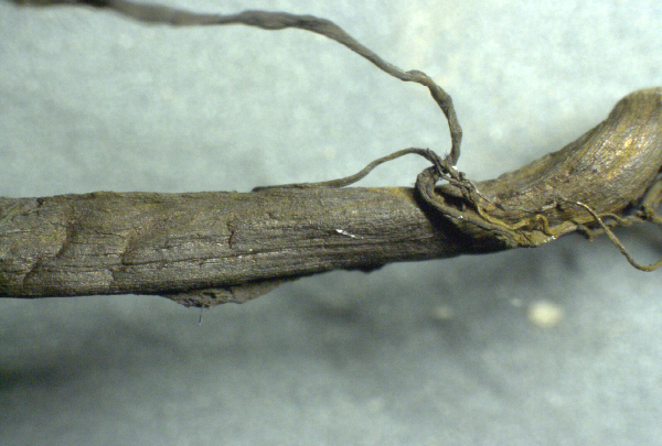
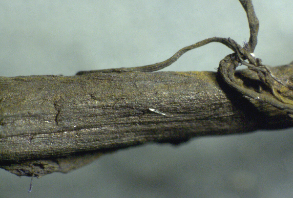
These clots are often referred to as "blood clots" but they are nothing at all like normal clots, and they consist of far more than mere blood cells. Unlike normal clots which are gelatinous, almost jelly-like, these so-called "clots" contain extremely large, complex, repeating structural elements (all shown below) that are clearly being constructed in the blood of the victims who died from these clots.
All of these clots were extracted from patients within a few hours of their death. These are not the result of post-mortem blood stasis. These are structures found in blood vessels and arteries. They are not congealed blood.
We wish to publicly thank Dr. Jane Ruby for connecting us to the embalmer (Richard Hirschman) who provided these clots. (Telegram channel T.ME/DRJANERUBY) Without the persistence of Dr. Ruby, you would not be seeing this report. Dr. Ruby is frequently featured on the Stew Peters Show (StewPeters.TV) and will also be my featured guest Monday on the Infowars.com broadcast.
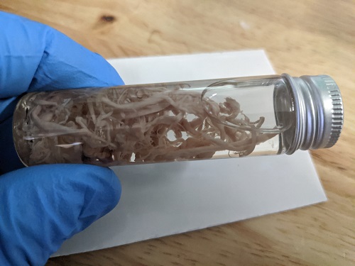
Here's a vial of these raw clots, washed of blood and preserved, before staining:
These structures exhibit the following shocking properties:
- They are tough, fibrous and resilient, showing material properties similar to small rubber bands.
- They consist of many strands of small, fibrous strands.
- These fibrous strands (see the very last photo set below) show repeating patterns of scale-like engineering, as if the body has been programmed to build another life form inside the blood vessels.
- There are strange crystalline-like structures found on these clots, exhibiting transparency and resistance to normal gram staining techniques.
- Below, you will find one example of a structure that appears to resemble a silicon-like biocircuitry or microchip-like structure. We don't yet know what it is.
- One of the photo sets below reveals what appears to be a biocircuitry wire which clearly shows repeating patterns and nano-scale interface structures that are assembled in a specific geometry for an unknown purpose.

Context for the photos you are about to see:
- I received these "blood clot" samples from a reputable embalmer (Richard Hirschman) who is active in the field of embalming and who confirmed these are not blood vessels or other tissues of any kind. They are structures that were evacuated from inside blood vessels during embalming procedures.
- I stained these samples using standard gram staining techniques used for microbiology in order to enhance structural contrast during microscopy. One of the samples below -- the more yellowish sample -- was stained only with iodine, not any violet-colored stains.
- The samples were then washed with ethyl alcohol and prepared on slides using standard tissue sample preparation for microscopy.
- Microscope magnification varies from 20x to 1500x, depending on the photo shown below. Magnifications are indicated with each photo set.
- I retain possession of these samples and can reproduce these photographs if required. Any competent lab microscopy operator could reproduce these photos using the same samples.
- My descriptions shown below are merely my own observations and are not intended to indicate certainty of the substances being identified. For example, when I talk about "biocircuitry" or "nanowires," I cannot confirm these are structures actually engineered for purposes of biocircuits. Merely, they resemble structures that seem to indicate such a purpose, but further research would be needed to confirm these observations.
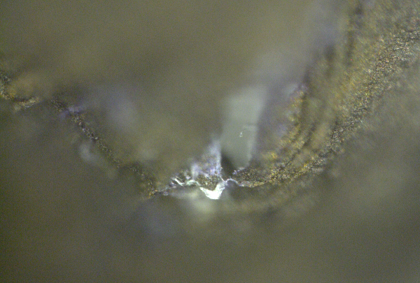
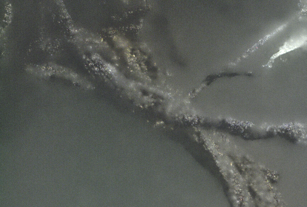
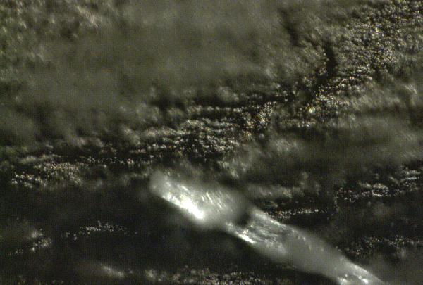
Microscopy photo set #1: Strange crystal-like nanostructures
This first set shows strange crystal-like structures that resist staining techniques and appear to show some sort of nano-scale, clear crystalline structures which would normally never appear in blood or blood clots.
Everything you are looking at in these photos is part of a blood clot extracted from an expired human being.
Magnifications shown here are 20x, 50x, 200x and 500x:

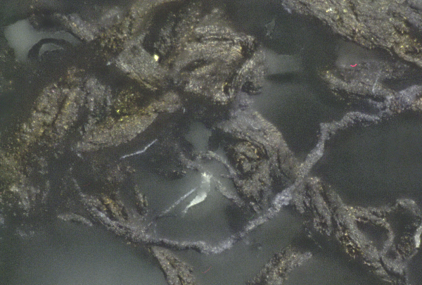
Microscopy photo set #2: Structures, strands and particles
This second set shows very close-up details on the strands, structures and particles found in these blood clots.
Magnifications shown are 20x, 50x, 100x, 200x, 500x, 1000x: (extreme magnifications causes a loss of depth of field which is why the highly-magnified photos seem so blurry in certain areas)
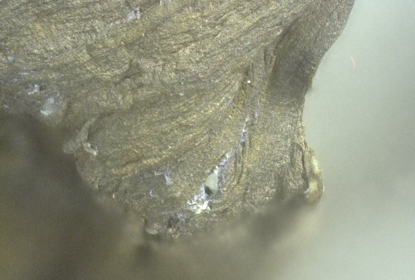
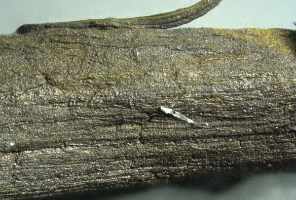
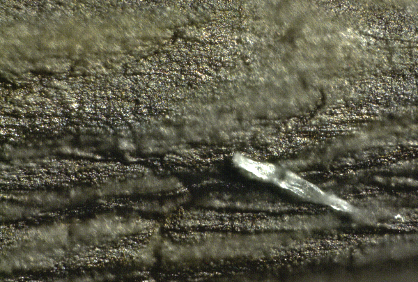
Microscopy photo set #3: Crystal-shaped structures
Crystal-like structures are attached to the bark-like structure of the blood clot. Remember, this clot is stained using a violet stain, which accounts for its dark purple color.
Magnifications are 20x, 50x, 100x, 200x, 500x and 1000x:
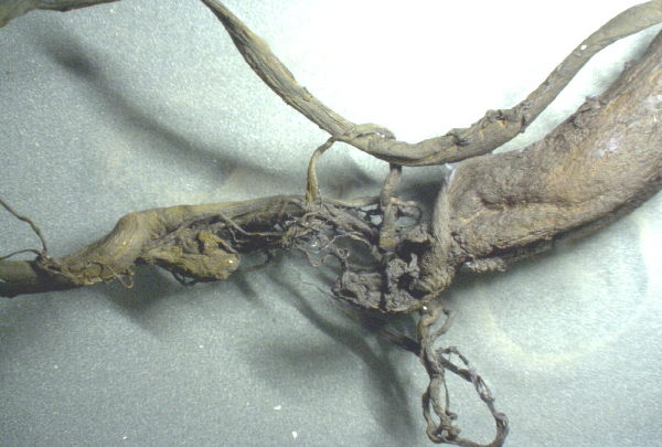
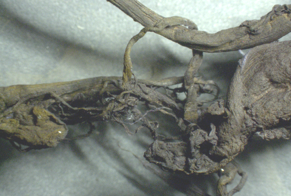
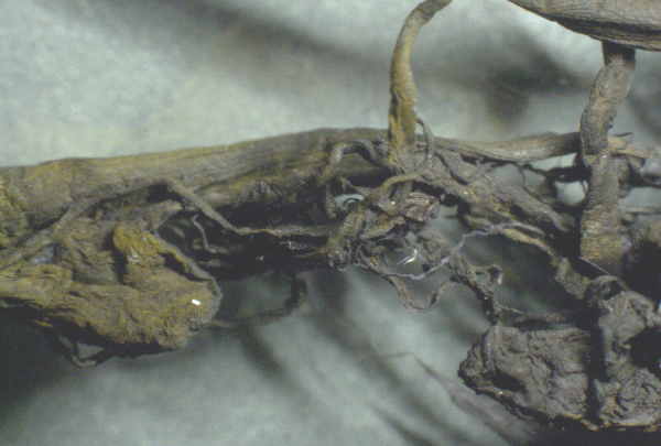
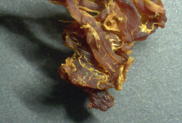
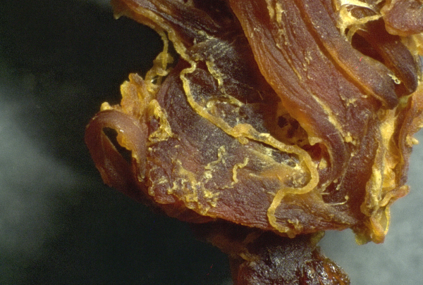
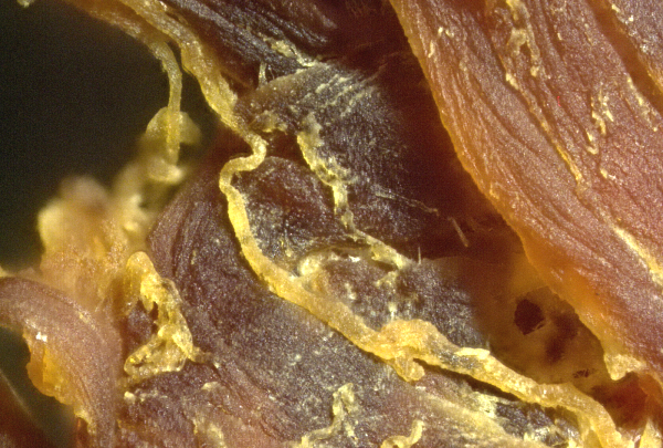
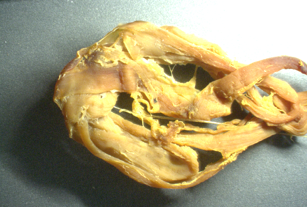
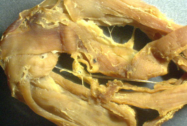
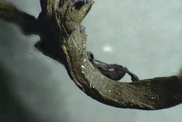
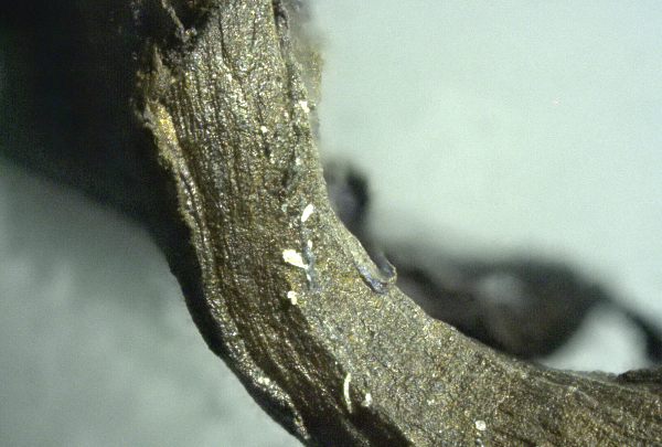
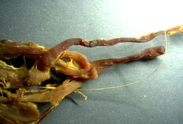
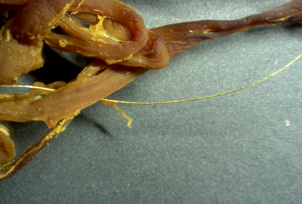
Microscopy photo set #4: Fibrous material is not simply congealed blood cells
The following sample was stained with iodine, then washed with ethyl alcohol. If you did not realize where this came from, you might think this was a sample of beef jerky or a chicken nugget. In reality, all of this is clot tissue that was found inside blood vessels or arteries.
As you can see, these are in no way "normal" blood clots. These have structure and are fibrous. They are clearly being built by the body, using protein synthesis instructions to create this large mass that nearly resembles muscle tissue. Yet it is being built inside the blood vessels.
Magnifications are 20x, 50x, 100x and 200x:
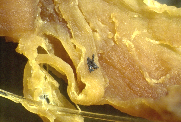
Microscopy photo set #5: Silicon-like "chip" structure
This series shows something that appears to resemble silicon-based microchip structures, although I cannot claim with certainty that this is a circuit of any kind. It simply resembles what micro-circuitry looks like at similar magnifications.
Magnifications used here at 20x, 50x, 100x, 200x and 500x:
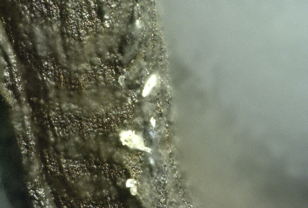
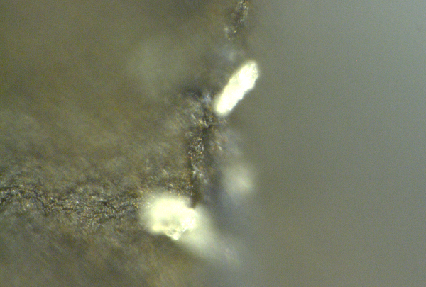
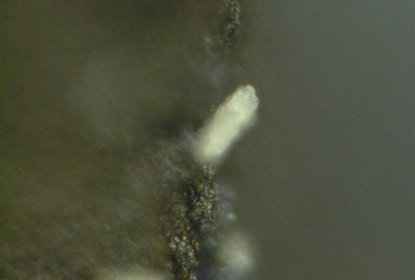
Microscopy photo set#6: Chalk-like white particles
An embalmer told me that blood emptied from the bodies of these people during embalming often appears to show "chalk-like" white particles which are visible even to the naked eye in certain cases.
My microscopy photos seem to have captured some of these chalk-like white particles which resist staining and seem to be scattered across certain regions of these clots.
Magnifications used here are 20x, 50x, 100x, 200x, 500x, 1000x and 1500x:
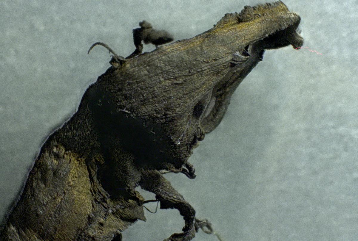
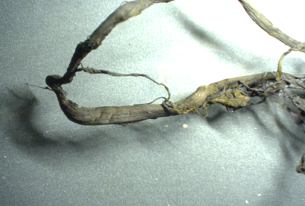
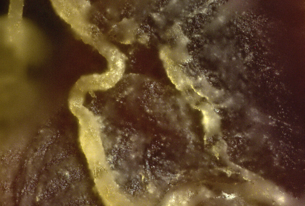
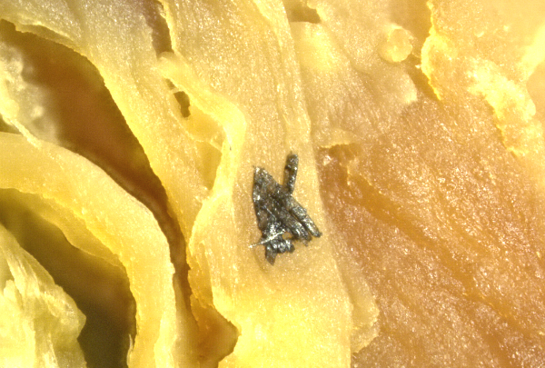
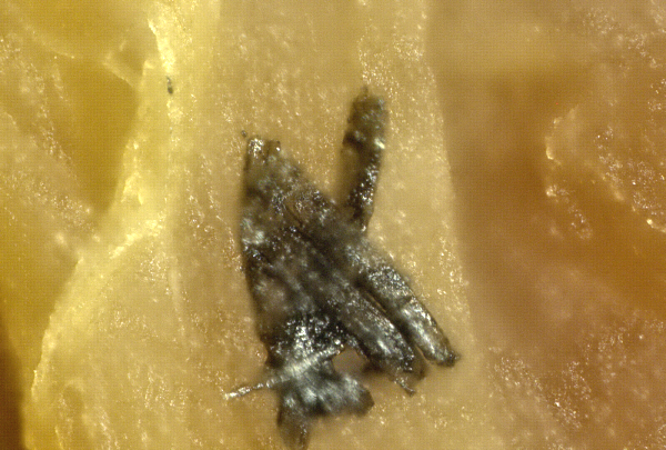
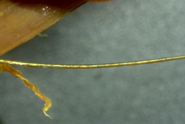
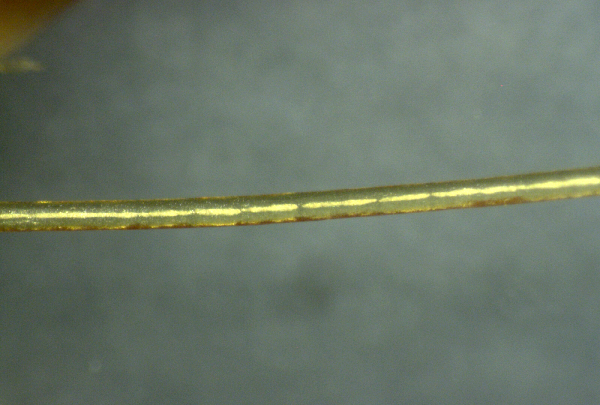
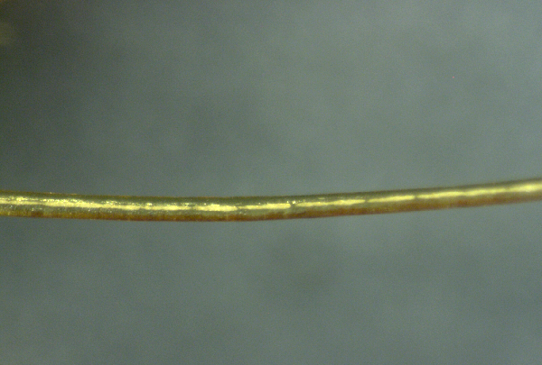
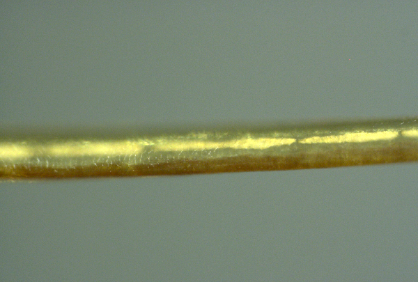
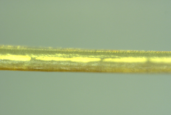
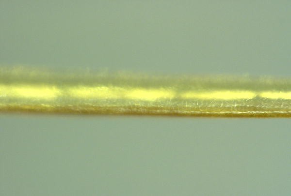
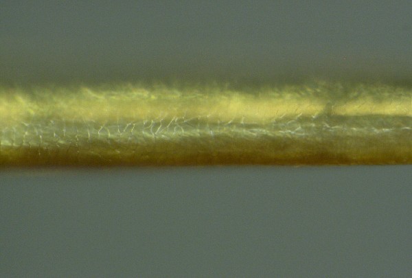
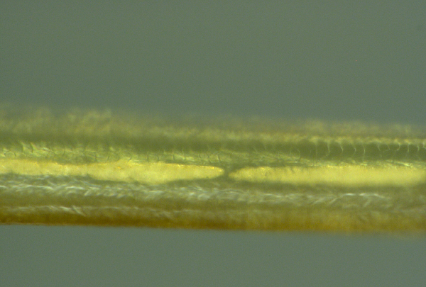
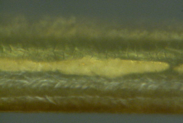
Microscopy photo set#7: "Nanowire" structures and repeating, structural scales
What follows here is a stunning look at what appears to be, at first, a micro-scale wire. Zooming it, we see a series of repeating structures along the top that appear to be nano-scale wire interface junctions. The entire "wire" is made of repeating segments, and its outer layer is covered in repeating "scale-like" patterns that actually resemble reptile skin more than anything human.
For the record, we don't know what these structures are. However, it's clear this doesn't belong anywhere in the circulatory system.
Finally, this fiber is not simply a human hair. It is firmly attached to the blood clot and when I tried to remove it, it would not tear away easily. This is not a contamination issue, it is a structure emanating from the clot itself. Everything you see here came out of a human being's blood vessels:
Magnifications used here are 20x, 50x, 100x, 200x, 200x, 500x, 500x, 500x, 1000x and 1500x:
What is all this?
We don't yet know what all these structures are. We know what they are not, however: They are not simply clotted blood cells. If they were, then at the 1500x magnification shown in the last photo, above, we would be able to see individual blood cells. These are not blood cells, they are protein structures.
Protein structures circulating in the blood like this, building up over time, are clearly being constructed by the body's cells. The ribosomes in the cells instruct the body what proteins to construct. These ribosomes are hijacked by mRNA gene therapy injections, which overwrite new instructions to the cells, causing them to manufacture something other than human.
I believe the structures you are seeing above are the result of mRNA protein synthesis instructions which have been injected into people under the false umbrella of "vaccines." I welcome input from other experts who may have other theories or explanations of where this is coming from.
More research is needed to confirm the function and composition of these structures, yet because of the extreme censorship and "science authoritarianism" that now exists in the world, no lab or university will dare examine these clots and honestly report the results. To do so would risk losing all NIH funding and federal grants, since the very same people who engineer vaccines and bioweapons also control most science funding in America.
Thus, only independent scientists, labs and journalists will dare tell the truth about these clots.
In conclusion, they are not "blood" clots. They are structures in the blood. They are "structural clots" or "fibrous clots" that are extremely large and are being constructed inside the body over time.
My grave concern is that every person who has been injected with mRNA instructions may be constructing these fibrous structures inside their bodies at this very minute, and that it's only a matter of time before they block major arteries or cause heart attacks, strokes or other acute causes of "Sudden Adult Death Syndrome" (SADS).
I believe these structures may very well explain why so many seemingly healthy adults are suddenly dying.
Hear more details on Brighteon.com and Infowars.com
I will be discussing these findings in more detail Monday mid-day on my Situation Update podcast at my channel on Brighteon.com:
https://www.brighteon.com/channels/hrreport
In addition, I am unveiling all these photos and findings as I host the Infowars.com "Alex Jones Show" broadcast Monday, June 13th, beginning at 11 am central. The three-hour show will feature several expert guests who will comment on these findings and present their own information about what these may be and how many people are being affected right now.
Tune in at www.InfoWars.com
And pray for humanity.
Mike Adams (aka the "Health Ranger") is the founding editor of NaturalNews.com, a best selling author (#1 best selling science book on Amazon.com called "Food Forensics"), an environmental scientist, a patent holder for a cesium radioactive isotope elimination invention, a multiple award winner for outstanding journalism, a science news publisher and influential commentator on topics ranging from science and medicine to culture and politics.
Mike Adams also serves as the lab science director of an internationally accredited (ISO 17025) analytical laboratory known as CWC Labs. There, he was awarded a Certificate of Excellence for achieving extremely high accuracy in the analysis of toxic elements in unknown water samples using ICP-MS instrumentation.
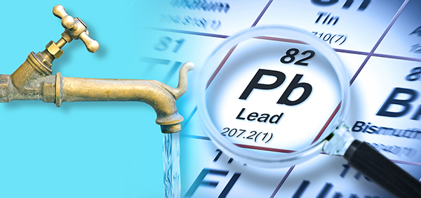
In his laboratory research, Adams has made numerous food safety breakthroughs such as revealing rice protein products imported from Asia to be contaminated with toxic heavy metals like lead, cadmium and tungsten. Adams was the first food science researcher to document high levels of tungsten in superfoods. He also discovered over 11 ppm lead in imported mangosteen powder, and led an industry-wide voluntary agreement to limit heavy metals in rice protein products.
Adams has also helped defend the rights of home gardeners and protect the medical freedom rights of parents. Adams is widely recognized to have made a remarkable global impact on issues like GMOs, vaccines, nutrition therapies, human consciousness.


Please contact us for more information.

