


New technique uses 25 times less radiation to produce tumor images in high-resolution 3D
Wednesday, April 09, 2014 by: David Gutierrez, staff writer
Tags: breast cancer, tumor images, radiation
- These 34 dentists reveal suppressed breakthroughs in holistic dentistry and oral health
- Codex Designates GMOs as Contaminants in Food
- Biotech company needlessly uses aborted human fetal cells to test artificial food flavors
- Alkaline diet food secrets revealed
- 11 common health symptoms hint at global depopulation 'slow kill'
- HuffPost: GMOs have never been proven safe
- Royal Rife: Cancer Cure Genius Silenced by Medical Mafia
- Cinnamon, Ginger and Onions Strongly Protect Us from Colds and Flu
- Food safety bill invokes Codex harmonization and grants FDA authority to police food safety of foreign nations
- Monsanto pushes bizarre conspiracy theory to deflect blame for GE wheat contamination of commercial crops
- The MMR vaccine, autism connection
- Ten surprising facts you didn't know about Donald Trump and the Health Ranger: Marrying immigrants, fighting for truth and expressing deep compassion for fellow human beings
- Indian black salve: The magical cancer cure
- The United Nations 2030 Agenda decoded: It's a blueprint for the global enslavement of humanity under the boot of corporate masters
- Study that sought to disparage health of eggs was authored by scientists with financial ties to Big Pharma
- Codex Threatens Health of Billions
- Sickening: Major food corporations use tissue from aborted babies to manufacture flavor additives in processed foods
- BREAKTHROUGH: The food you eat determines which genes get activated or suppressed, controlling disease vs. health throughout your life (the Health Ranger was right!)
- These 34 dentists reveal suppressed breakthroughs in holistic dentistry and oral health
- Chemtrails: Learn how to protect yourself from these treacherous poisons
- Royal Rife: Cancer Cure Genius Silenced by Medical Mafia
- Is your shampoo harming your health?
- Green tea extract may help prevent type-2 diabetes through improved glucose tolerance
- 11 common health symptoms hint at global depopulation 'slow kill'
- How to Use Organic Neem Leaf for Flea and Tick Control for Cats
- The best and worst forms of magnesium to take as a supplement
- Biotech company needlessly uses aborted human fetal cells to test artificial food flavors
- Indian black salve: The magical cancer cure
- Alkaline diet food secrets revealed
- Zeolite truths, myths, benefits and exaggerations... explosive interview with zeolite expert blows the lid on what's REAL vs. BOGUS
- The United Nations 2030 Agenda decoded: It's a blueprint for the global enslavement of humanity under the boot of corporate masters
- Toddler injected with 37 vaccines before the age of two left paralyzed and wheelchair-bound for life
- Folic acid proven to prevent heart disease and stroke in study
- Flu vaccines causing massive spike in deaths of elderly across the UK, warn health officials
- Heal Yourself in 15 Days by Cleaning Up Your Skin Exposure (Part Ten)
- The Great Culling has begun: Will your genetic lineage survive?
- Vaccine mafia earns 'F' in science: Australian Health Minister utters the most insanely stupid anti-science statement ever recorded... 'no risks in vaccinating children'
- The best and worst forms of magnesium to take as a supplement
- The United Nations 2030 Agenda decoded: It's a blueprint for the global enslavement of humanity under the boot of corporate masters
- Colorado Batman shooting shows obvious signs of being staged
- Vaccinated children have up to 500% more disease than unvaccinated children
- NASA admits to spraying Americans with poisonous chemtrails
- What's really in vaccines? Proof of MSG, formaldehyde, aluminum and mercury
- The real vaccine scam is not that they cause autism, but that they don't even work!
- The MMR vaccine, autism connection
- New NASA research points to possible HAARP connection in Japan earthquake, tsunami
- Green tea extract may help prevent type-2 diabetes through improved glucose tolerance
- Reverse and eliminate cataracts naturally without surgery
- Beat cancer with 35% hydrogen peroxide
- NASA gives thumbs up to use of colloidal silver as antibiotic in space; FDA has no jurisdiction in high orbit
- Bicarbonate of Soda Used to Cure Stage Four Prostate Cancer
- Inflammation is the cause of nearly all disease - Here's how to prevent it
- Anti-foaming agent found in Chicken McNuggets
- Royal Rife: Cancer Cure Genius Silenced by Medical Mafia
- The Coming Gold Revaluation: Strategic Financial Realignment in an Era of Dollar Collapse
- The AI Data Center Wars Have Begun… Farms, Water and Electricity is Stripped from Humans to Power the Machines
- Urgent Wake-Up Call: The Coming AI Robot Wars and the Great Human Unity
- Eleven days before Iran bombed Tel Aviv, my microscope revealed haunting images of EXACTLY what would happen
- The War on Light: How Governments and Big Pharma Keep You Sick By Blocking Healing Photons
- THE AI RACE IS ALREADY WON: How China’s power dominance (and America’s climate lunacy surrender) secured its victory in the race to AI superintelligence
- Why the U.S. Government May be Seeking to Slaughter 200 Million Americans to Free Up Excess Power for AI Data Centers and the Race to Superintelligence
- Trump's Trojan Horse: How Stablecoins Are Secretly Paving the Way for a CBDC Control Grid – an Interview with Catherine Austin Fitts
- DECENTRALIZED SPIRITUALITY and the true teachings of Christ: Overcoming the censorship, threats and lies of organized religion to truly know God and the Universal Christ
- EXCLUSIVE REPORT: The Trump-Putin Meeting in Alaska - A Historic Pivot that Redefined Global Power
- Morphic resonance “remote viewing” reveals iconic Middle East images of stealth bombers, a falcon and a one-horned ram
- 18 MILLION COVID-JABBED Japanese folks shown to have SIGNIFICANTLY HIGHER DEATH RATES during first year after injection with mRNA clot shots
- AI & economic liberty: Will decentralized tech save human autonomy?
- Franken-butter: Startup backed by Bill Gates creates butter from thin air
- HEALTH SECRETS: How to Instantly Block MSG Toxicity Using Natural Substances (and the secret of Methylene Blue)
- BOMBSHELL: DNA testing kits are a SCAM to develop ethnic-specific bioweapons
- BOMBSHELL: Internal Pfizer documents exposed and reveal at least 16 PERCENT of their mRNA vaccine "adverse events" are REPRODUCTIVE DISORDERS
- How a nasal spray vaccine could turn Americans into walking bioweapons this Fall


"This new technique can open up the doors to the clinical use of computed tomography [CT] in the breast diagnosis, which would be a powerful tool to fight even better and earlier against breast cancer," said Maximilian Reiser of the Ludwig Maximilians University in Munich (LMU), one of the researchers who helped develop the technique.
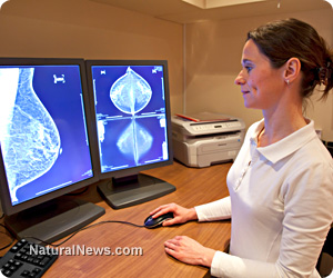
Early detection is linked to significantly improved breast cancer outcomes. Currently, the standard breast cancer screening method is "dual-view digital mammography," which uses radiation to take two separate images of the breast. Because these mammograms render a three-dimensional organ in only two dimensions, they typically fail to detect 10 to 20 percent of tumors. In addition, 2D mammograms often render false positives, as normal tissue can take on a strange appearance when flattened into two dimensions.
Although 3D CT scans produce much-higher-resolution images than 2D mammograms, they typically require much more radiation to do so. This prevents their use in routine cancer screening -- particularly for highly radiation-sensitive organs such as the breast.
Advanced techniques use less radiation
The new, lower radiation CT scan was tested in a study published in the Proceedings of the National Academy of Sciences in October 2012. The technique was developed by physicists, radiologists and mathematicians from LMU, the European Synchrotron Radiation Facility, and the University of California-Los Angeles.The researchers achieved such a dramatically lower radiation dose by changing three separate factors of normal CT scans. The first change involved replacing standard X-rays with high-energy X-rays which are more likely to pass through tissue and thereby expose the body to six times less radiation. The second change involved a technique called "phase contrast imaging," which requires fewer X-rays to produce the same image. The third change involved reducing the number of X-rays even further and then applying a mathematical algorithm called "equally sloped tomography" (EST) to reconstruct a higher-resolution image.
In the 2012 study, the researchers successfully used the EST algorithm to produce a high-resolution, 3D image of the breast using 25 times less radiation than a mammogram. They then took images of the same breast using several other standard 3D imaging techniques. In a blind test, five independent LMU radiologists all ranked the images produced from the new technique as having the best contrast, sharpness and image quality.
"Three-dimensional reconstructions, like the ones created in this research, are produced using sophisticated software and a powerful computer that can combine many images into one 3-D image, much like the slices of an orange," UCLA researcher Jianwei (John) Miao said. "By rethinking the mathematic equations of the software in use today, we developed a more powerful algorithm that requires fewer slices to get a clearer 3-D picture."
Applications still far off
"After dramatically reducing the dose delivered during the examination of the breast, our next objective is to develop this technique in the early visualisation of other human diseases and to work towards its clinical implementation," said researcher Paola Coan.Unfortunately, it may be some time before the new technique is ready for regular use. One of the major hurdles is the need to produce an X-ray source small enough to be practical at hospitals.
"Many research groups are actively working to develop this device and once this hurdle is cleared, the new X-ray technique is poised to make a big impact on society," researcher Emmanuel Brun said.
Sources for this article include:
http://www.esrf.eu
http://www.esrf.eu
http://science.naturalnews.com


Breast cancer at FETCH.news
Get independent news alerts on natural cures, food lab tests, cannabis medicine, science, robotics, drones, privacy and more.

Take Action: Support Natural News by linking to this article from your website
Permalink to this article:
Embed article link: (copy HTML code below):
Reprinting this article:
Non-commercial use OK, cite NaturalNews.com with clickable link.
Follow Natural News on Facebook, Twitter, Google Plus, and Pinterest

Science News & Studies

Medicine News and Information

Food News & Studies

Health News & Studies

Herbs News & Information

Pollution News & Studies

Cancer News & Studies

Climate News & Studies

Survival News & Information

Gear News & Information

News covering technology, stocks, hackers, and more


"Big Tech and mainstream media are constantly trying to silence the independent voices that dare to bring you the truth about toxic food ingredients, dangerous medications and the failed, fraudulent science of the profit-driven medical establishment.
Email is one of the best ways to make sure you stay informed, without the censorship of the tech giants (Google, Apple, Facebook, Twitter, YouTube, etc.). Stay informed and you'll even likely learn information that may help save your own life."
–The Health Ranger, Mike Adams